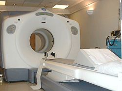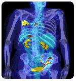
Positron emission tomography (PET imaging or PET scan) is nuclear medicine imaging with an injected or swallowed radioactive isotope (radiopharmaceutical). A PET scan measures body functions, such as blood flow, oxygen use and sugar (glucose) metabolism. These scans help doctors evaluate organ and tissue functions. Nuclear medicine imaging studies are less directed toward picturing anatomy and structure, and more concerned with depicting physiologic processes within the body, such as rates of metabolism or levels of chemical activities. Areas of greater intensity -- "hot spots" -- indicate where large amounts of the radiopharmaceutical have accumulated resulting in high levels of chemical activity. Less intense areas -- "cold spots" -- indicate a smaller concentration of radiopharmaceutical and less chemical activity.
The combined PET/CT scans provide images that pinpoint abnormal metabolic activity within the body. The combined scans provide more accurate diagnoses than the two scans performed separately.
PET/CT scans:
- Detect cancer
- Determine the spread of cancer
- Assess the effectiveness of treatment
CT Procedure with Contrast

If you had a CT scan procedure with IV contrast (dye), contact your physician if any unusual symptoms occur, such as a rash or hives. If your IV site is sore, reddened, or swollen, apply a warm, wet washcloth to the area for 15-20 minutes four times a day and elevate your arm on a pillow. Call your physician if symptoms persist for more than 48 hours.
If you had a CT scan procedure with oral contrast (barium), drink extra fluids to aid in passing the contrast from your system. If you become constipated, contact your physician.
If any life-threatening symptoms occur, go to the Alton Memorial Emergency Department or call 911.
Additional information
To schedule an appointment with the Alton Memorial Hospital Imaging Center, please call
618.463.7647 or
email us.